Poltava, prospekt Vitaliia Hrytsaienka 9 Weekdays: 8:30-15:00, Weekend: weekend +38 (0532) 56-02-11+38 (095) 688 25 07
Features of breast diagnosis before and after plastic surgery
 Any surgical intervention requires a preliminary examination. This makes it possible to correctly assess the patient's condition and avoid possible negative situations or consequences during operations. Mammoplasty is no exception. Also, a large number of questions arise about the reliability of breast examinations in the presence of an implant.
Breast examination before mammoplasty
The preparatory process includes not only a standard package of tests (total urine, blood, sugar, cardiogram, fluorography, etc.), but also a mandatory examination of the breast using ultrasound and x-rays. It includes:
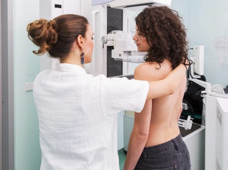
- X-ray of the UCP;
- Ultrasound of the mammary glands and lymph nodes.
 Such a diagnosis is of great importance. Indeed, during its conduct, problems or diseases may appear that the patient herself is not aware of. It is primarily about mastopathy and various tumors. If a neoplasm is detected in the chest and timely access to oncologists can cure cancer. Especially if it is at an early stage of its development.
If problems are discovered during the preparation process, the operation is naturally delayed, and it is recommended that the client contact the appropriate specialists.
Breast Diagnosis with Implants
The main diagnostic methods for breast cancer, with or without implants, are ultrasound. Additional examination methods are prescribed only if the study reveals formations of a suspicious nature. Sometimes even computed tomography is prescribed. But it is prescribed only as a last resort, if the previous results do not make it possible to draw an unambiguous conclusion about the disease.
The examination is not affected by the implant placement method. In plastic surgery, they are used two:
- under the mammary gland;
- under the muscle.
 The latter method, according to experts, can cause certain problems. The fact is that when penetrating through the armpit, lymph flows can be affected. With a decrease in size (reduction), the formation of lipogranulomas is even possible. This is a neoplasm, but it consists of adipose tissue that is dead. Do not confuse it with a lipoma. This is a benign tumor from living tissue cells that have undergone a modification. Lipogranulomas appear due to surgery, in places of scarring. If an ultrasound is performed by a mammologist with experience, then in any case he will pay attention to education and advise an additional examination. A puncture clearly gives the opportunity to talk about the presence or absence of cancer.
Do implants interfere with breast diagnosis
The presence of implants makes ultrasound examination of the breast somewhat difficult. But for an experienced mammologist with special equipment, this is not a difficulty.
After an operation to correct the mammary glands, an annual ultrasound is recommended. This allows you to closely monitor the condition of the implant, to eliminate its rupture or leakage, to avoid inflammatory processes. With ultrasound, the presence of a prosthesis does not cause any complications. But with mammography, various options are possible. When installed on the pectoral muscle, a part of the gland is blocked, which makes it impossible to see the full picture. But in this case, you can apply other methods that allow you to see the full picture. The introduction of an implant under the muscle reduces the closed area.
specialists of the Clinic for Plastic Surgery
27-12-2019
Similar news
