Poltava, prospekt Vitaliia Hrytsaienka 9 Weekdays: 8:30-15:00, Weekend: weekend +38 (0532) 56-02-11+38 (095) 688 25 07
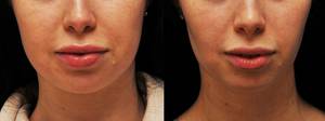
Resection of the fatty body of the cheek (removal of the lumps of Bisha)
Procedure of Bichat’s fat body removal (Excerpt from the I.Ju. Slusarev book called "The facelifting")
One way to correct lateral surface contour is dosed resection of fat body of cheek spurs (corpus adiposum buccae), the most interfacial and self-contained of which are Bichat’s fat bodies..
A result of the procedure is the emphasizing of the zygomatic region lower contour by reducing the volume of the cheeks.
It is more common that fat resection is performed not as a stand-alone operation, but it complements the various methods of open face lifting. This is due to ease of access to the fat body in SMAS Sub-plane.
Resection of Bichat’s fat bodies
with access through the buccal mucosa
It is often made at a young age and is aimed to make a proper relief of the malar area..
The surgery starts with finding the anatomical landmarks in the oral cavity. It is easy to find the front edge of the masseter muscle by putting the finger in the mouth. The excretory duct of the salivary gland can be found is a slightly medial direction. The location of intended buccal mucosa incision is being marked 1 cm deeper from the excretory duct and 1 cm away from the upper mucobuccal fold.
It is inappropriate to anesthetize a large area when removing the fat bodies. It is enough to inject 1.5-2 ml of anesthetic solution in the incision area that is under the mucous membrane. Then the incision of mucosa up to 1 cm along the mucobuccal fold is being performed with pointed scissors or a scalpel. M. buccinator,fibers, which are located directly under the mucosa, are being expanded blankly with the pointed clamp. While pressing a finger onto the outer surface of the cheek and simultaneously folding it back, it is possible to visualize the fat body at the wound.
While pulling and grabbing the fat body gently with forceps, it is easy to withdraw its entire volume in the mouth cavity. It is highly unadvisable to pull it strongly and make significant efforts to remove it. This can lead to disruption of the capsule integrity and may cause bleeding.
Trimming the fat body in the neck is being performed after the preliminary legation or coagulation of its foundation The mucous membrane is underrun together with buccinator and is tied with one seam. The fast absorbable sutures are best for this purpose.
After performing the melonoplasty, it is necessary to apply the removed fat bodies to the cheek and photograph them.
Transferring the Bichat’s fat bodies
Transferring the Bichat’s fat bodies can be done both independently as well as it may complement lifting procedures. By moving the fat body volume, a more expressed correction of midface, the desired voluminosity of malar area and relieve of the lower cheek section can be achieved.
After performing the buccal mucosa anesthesia, the incision is made along the mucobuccal fold; it should be no longer than 1.5 cm. The main landmark is the excretory duct of the salivary gland. The incision should be located 1 cm behind it, and 1 cm away from the upper transitional fold. By expanding the tissues with a clamp blankly and pressing with a finger to the outer surface of the cheek, the part of the fat spur is placed in the wound. Do not try to remove a large volume; it is enough to place a diameter of 1 cm out of the wound.. This is usually enough to underrun this area crosswise and immerse it into place. The free ends of the suture are placed in the direction of the tongue.
There are two different ways to transfer the Bichat’s fat body. In the first case it is moved to a given position under the bucco-zygomatic fat. To do this, it is necessary to make a tunnel from incision performed under the fat block, in the direction of the zygomatic bone tuber. Since tunneling is performed without visual control, care should be taken not to damage the cheek muscles. The greater zygomatic muscle must stay at the bottom of the formed tunnel. The muscles are connected quite intimately with the fat package located on them, so the separation of this layer should be carried out by blunt scissors jaws separation with minimal force.
The second option involves the formation of subperiosteal tissue tunnel and pulling of fat body to a given region through it.
This option allows to concentrate the main volume at the specified location. The formed subperiosteal cavity is only the transfer tunnel for the fat body. It contains a smaller part of it, and the main volume is concentrated under soft layers.
To do this, the anesthetic solution is injected under the periosteum over the lateral part of the zygomatic bone through the incision in the mucous membrane already made. The tissues are expanded immediately prior to the periost. Then it is dissected in frontal projection of the m. master tendon. Using the raspatory, the periosteum is lifted slightly in the lateral direction to the zygomatic bone tuber and is cut with the width of up to 1 cm at this point. The main volume of Bichat’s fat bodies should be moved in this space. Width of subperiosteal tunnel should be 1-1.5 cm.. t is not desirable to approach the lower external edge of the arcula, since there is a risk of injury. r. zygomaticofacialis.
Moving and fixing of the fat body is performed in the following manner. Both free ends of the suture, which is used to underrun the fat body edge, are inserted into the long straight needles and are passed sequentially through the tunnel formed into the top section of the cavity. From that spot they pierce the skin at different angles and are taken to the surface at a distance of 1.5 cm below the edge of the lower external edge of the arcula from one point.
The edge of the fat body is captured with tweezers. It is possible to place it on the planned surface by pulling the two sutures on the outside and helping to move the fat body with tweezers at the same time.
Fixing the fat body in a predetermined position it is performed by fastening the suture ends. They can be fastened on the outside of the gauze swab. But it is quite inconvenient, since it is necessary to perform daily bandaging with control of pressure exerted by gauze sponge on the surface of the tissue. uture support should be performed within 5-6 days.. A shorter fixation can lead to a displacement of the fat body from the predetermined position.
It is more convenient to fasten the sutures together and immerse the knot under the skin through micro-incision. It is undesirable that the funnel shaped retraction would appear at the knot area. It is necessary to arrange the suture so that the contour of tissue would change in the form of the soft bend. Since the sutures with a short absorption period are being used for the fixation, this deformation will disappear by itself after a short time.
specialists of the Clinic for Plastic Surgery
21-09-2016


